


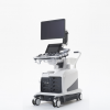
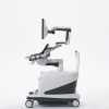
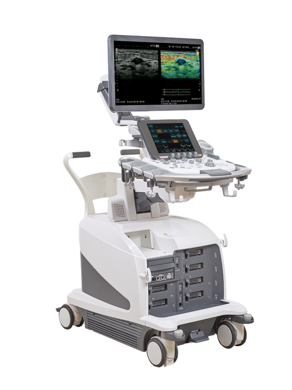
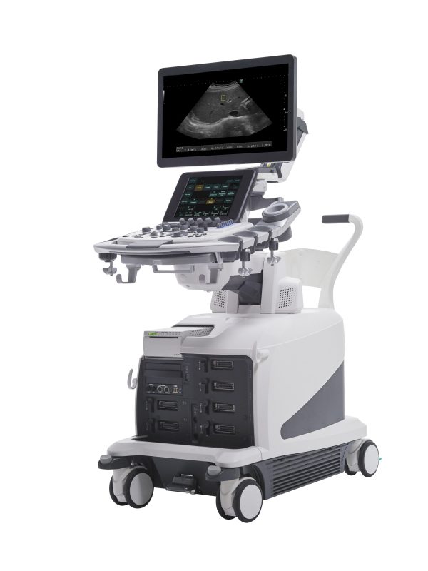
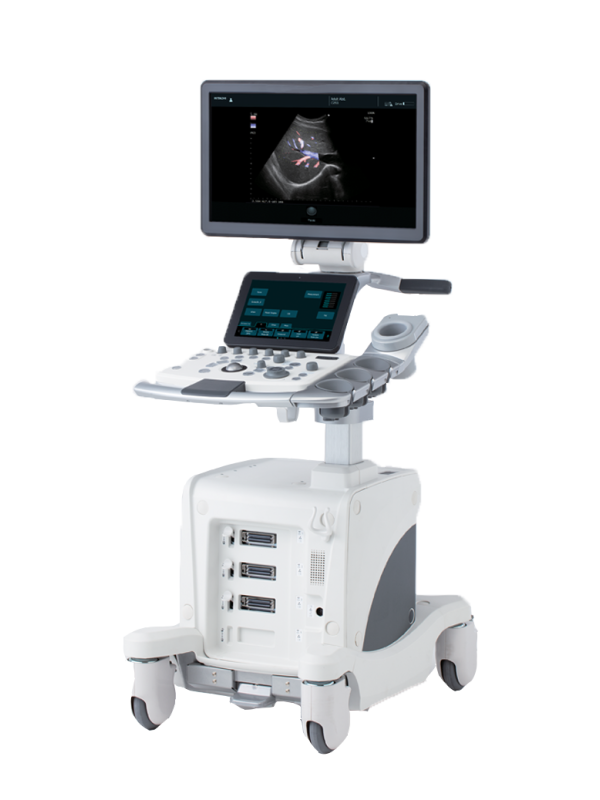
The LISENDO 880LE is FUJIFILM’s premium 3D/4D diagnostic ultrasound system for cardiologists in any clinical setting, designed to be a true one-system solution for adult, paediatric and fetal heart patients. The LISENDO 880LE platform is redefining the vision for cardiac ultrasound by providing exceptional clinical performance combined with state of-the-art analysis and features.
The ultrasound platform features:
- Pure Image to attain remarkable fundamental image quality in cardiology, achieving more reliability during diagnosis and treatment
- Your Application to reach a most sophisticated visualization of the blood flow patterns and its mechanisms as well as an impressive display of 4D cardiac image
- Seamless Workflow, ensuring high user operability by applying Artificial Intelligence (AI) technology to significantly improve examination efficiency
FEATURES
The LISENDO 880LE is FUJIFILM’s new premium 3D/4D diagnostic ultrasound solution for cardiologists that redefines the vision of cardiac ultrasound by providing exceptional clinical performance coupled with state-of-the-art features and analytics.
With our innovative cardiac imaging features, we are able to move hemodynamic evaluations to a new level:
New generation of cardio-vascular transducers, Phased Array single crystal transducers and Linear 4G CMUT (Capacitive Micro machined Ultrasound Transducer) with high quality imaging adapted for a variety of examinations.
Cardiac Transducers : the 3D and 4D phased array transducers have been newly designed with the last single crystal technology to realize the high spatial, temporal and contrast resolution especially required for cardiology. With an improved shape that is comfortable to hold and easily fits in intercostal spaces, it can reduce variable factors such as user skill- and patient disease-dependencies that can inhibit image clarity.
Vascular Transducers: Conventionally, the superior resolution obtained with high frequency ultrasound is only used for superficial examinations due to its limited penetration. Lower frequencies are needed to examine deeper structures, thus multiple transducers are required to cover the full range of vascular ultrasound examinations. The world’s first practical use of the CMUT silicon wafer technology was introduced by us in 2009. In the next-generation technology, thanks to exclusive FUJIFILM patents the bandwidth and sensitivity has been dramatically increased. In addition Pulsed Doppler and Color Doppler allowing to image blood flow as well as Harmonic mode are now available. CMUT now delivers a one-probe solution for a wide range of ultrasound examinations.
New transmission and reception technology eFocusing, resulting in high definition imaging from the near to far field.
The eFocusing transmission and reception technology newly developed for LISENDO 880LE significantly improves S/N and reduces focal dependency. The outstanding clarity of clinical images is performed from near to far field with excellent penetration at higher frequencies.
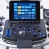
OLED monitor achieving high contrast resolution
LISENDO 880LE has adopted the latest technology, a 22″ OLED monitor, for an optimum image display. The self-luminous OLED displays true black so a previously unattainable contrast resolution can be achieved. It is the ideal monitor choice for diagnostic ultrasound, with the highest quality grayscale display.
HD ANALYTICS
Perfom Optimal Diagnosis for Heart Failure Diagnosis.
For unique and accurate assessment of cardiac hemodynamics, Hitachi has combined a collection of the most powerful analytical tools, packaged as HDAnalytics, for Lisendo 880.
Global Longitudinal Strain
GLS, the ratio of change in LV endocardium length, can be altered significantly in patients with heart failure, while maintaining a normal Ejection Fraction (EF).

eTRACKING with Wave Intensity
eTRACKING provides early detection of atherosclerosis, vessel wall stiffness independent of blood pressure, and estimation of the vessel age from the β value.
The Wave Intensity provides an index for the timing of early systole and end systole during the rapid change of pressure and velocity.

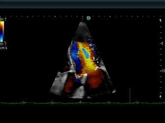
Virtual Contrast
Virtual Contrast is a non-invasive, high definition blood flow imaging mode that drastically improves sensitivity for the visualization of the endocardial border in the left ventricle; it is particularly useful for diagnosing technically difficult patients.
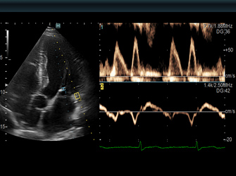
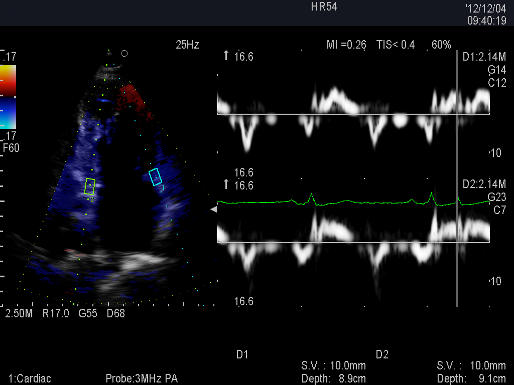
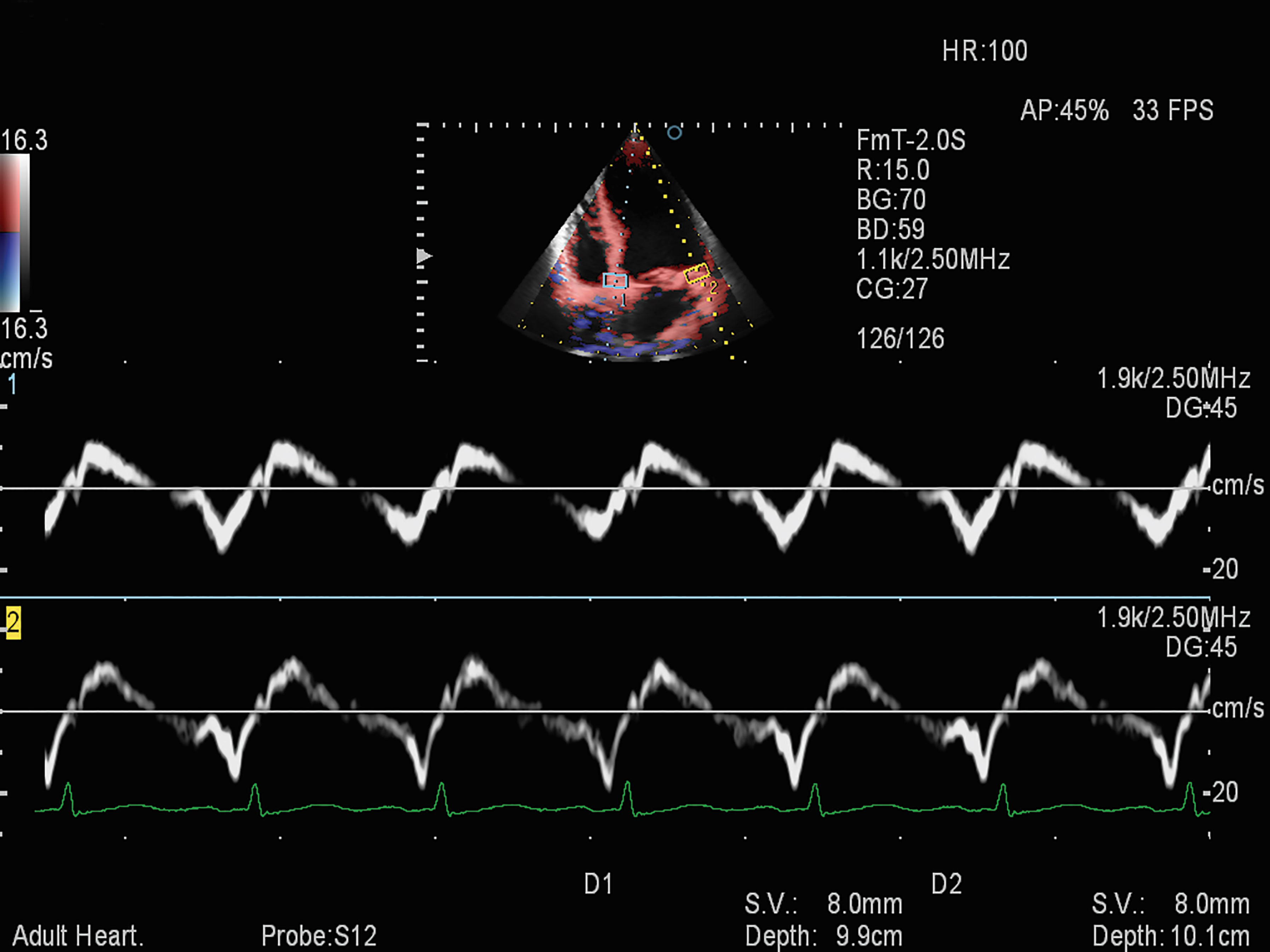
Dual Gate Doppler with R-R Navigation
Simultaneous FFT spectral analyses from two sample gates are provided by Dual Gate Doppler for effortless comparison.
In addition, R-R Navigation automatically detects appropriate heartbeats for measurement in patients with irregular heartbeats. Measurements such as the E/e’ ratio, isovolumetric contraction and relaxation times, as well as an evaluation of dyssynchrony in septal and lateral walls can be obtained from the same heartbeat.

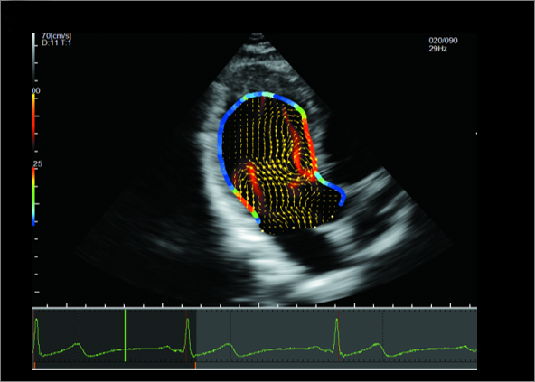
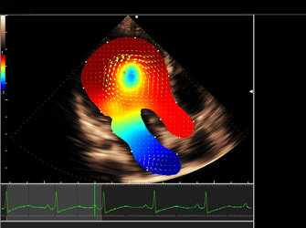
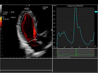
Vector Flow Mapping
VFM can display the resultant hemodynamic effects within the LV during systole of defects captured by GLS. The energy loss display maps the energy dissipated by flow disturbance providing possible early detection of heart failure from information obtained within the heart chamber.

Perfect with 2DTTE Transducer.
The phased array transducer has been designed to realize high spatial, temporal, and contrast resolution that is essential for cardiology.
Focused at all depths with eFocusing.
eFocusing, the newly developed transmission & reception technology realizes significant S/N improvement and reduces focal dependency whilst providing excellent penetration at higher frequencies.
With a one touch operation, eFocusing offers a real-time display unmodified by image processing.

Automated Cardiac Measurement. Smart & Precise.
With Hitachi’s Big Data and Artificial Intelligence (AI), high precision in automated measurements of complex cardiac functions can be attained.

Fractional Area Change

LV Volume (EF)

RA Volume

LA Volume

Automated ED-ES Dectection
Beyond Basic Hemodynamic Evaluations
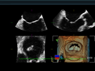
Dynamic 3D Cardiac
Lisendo 880 offers Bi-plane Imaging, 3D Zoom, Active 3D, and Wide-Angle 3D Live Imaging to provide a comprehensive set of data for your 3D and 4D Cardiac Evaluation and Analysis. The acquired 3D data can be used for different analysis packages, including valve diameter measurement, 3D morphological observation, volume calculation, and tracking.
Cardiac 3D
4D MV Assessment
3DTEE

4D LV Analysis
iEF
Automated calculation of Ejection Fraction from 3D volume data. The Biplane images (4ch and 2ch) are displayed with ED/ES frames selected automatically.

i2DTT
The fully automated speckle tracking function provides precise quantification of strain and strain rate for the left and right ventricles and left atrium for visualization and analysis of myocardial mechanics.


